[The industry's No. 1 journal on diagnostic imaging!]
Features on clinically relevant topics across the entire field of diagnostic imaging,
explaining everything from the basics to the latest information with beautiful photographs in an easy-to-understand manner!
Includes a quiz-style series and a participatory series that answers reader questions.
...Target audience: Radiology and related fields, including physicians and technologists.
[CONTENTS]
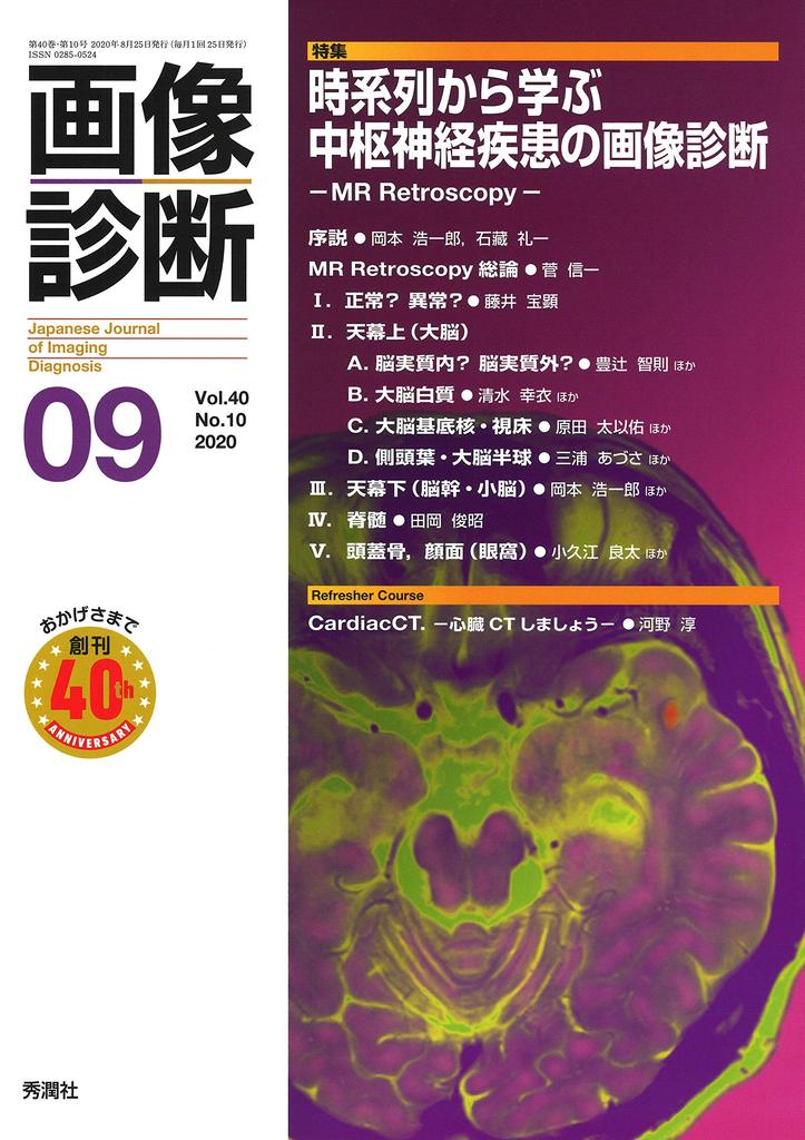
Learn about the timeline of central nervous system disease imaging using MR Retroscopy.
Introduction ● Koichiro Okamoto, Reiichi Ishikura
Introduction to MR Retroscopy ● Shinichi Suga
I. Normal? Abnormal 1. Headache in patients undergoing chemotherapy for malignant tumors ● Takaaki Fujii, Satoshi Terae
II. Cerebrum
A. Intracerebral or extracerebral parenchyma 1. Meningioma mimics - Interpreting images without preconceptions ● Tomonori Toyotsuji, Kentaro Akazawa, et al.
2. A mass connected to the olfactory tract in the midline of the forehead ● Minako Azuma, Yoshihito Kadota, et al.
B. Cerebral White Matter
1. Subcortical White Matter Lesion Showing a Growing Trend Over Several Years ● Shimizu Yukie, Harashima Toko, et al.
2. Cerebral White Matter Lesion - Is the FLAIR Hyperintensity Really Chronic Ischemic Change? ● Kojita Yasuyuki, Kanda Tomoki, et al.
3. Suspected Multiple Sclerosis - Is it Multiple Sclerosis? ● Kimura Yukio, Chiba Emiko, et al.
C. Basal Ganglia and Thalamus
1. Bilateral Basal Ganglia Abnormal Signals - Inflammation? Tumor? ● Harada Taisuke, Yamazaki Yasuyuki, et al.
2. Slowly Developing Bilateral Thalamic Lesions - Careful Follow-up of Pediatric Microlesions! ● Kitahara Sawako, Ito Ryuta, et al.
D. Temporal Lobe and Cerebral Hemisphere
1. Non-Improved Limbic Encephalitis - Mimicker ● Miura Azusa, Fujiwara Masahiro, et al.
2. Mass Lesion Appearing After Surgery for a Medial Temporal Lobe Lesion ● Shohei Inui, Ryo Kurokawa, et al.
3. A patient with a history of optic neuritis of unknown etiology six months prior presented with sudden onset of headache and right hemiparesis. ● Toshiaki Akashi, Shiho Sato
4. Widespread lesion in one cerebrum, including the brainstem. ● Minako Azuma
III. Subtentorial lesion (brainstem and cerebellum)
1. Solitary midbrain nodular lesion in a young adult - Demyelinating lesion? Inflammatory/granulomatous lesion? ● Koichiro Okamoto, Junichi Yoshimura
2. Is it really an artifact? A case of diplopia and right abducens nerve palsy during the progression of pulmonary sarcoidosis. ● Tomonori Toyotsuji, Hiroshi Miura, et al.
3. Midbrain mass lesion in an adult - A case with an unexpected course. ● Masahiro Fujiwara, Noriyuki Toyama, et al.
IV. Spinal cord
1. Cervical spinal cord deformation and abnormal signals. ● Toshiaki Taoka
2. A Case of Complex Pathology and Clinical Changes During Follow-Up of Cervical Myelopathy ● Tokumaru Aya, Saito Yuko et al.
V. Skull, Face (Orbit)
1. Subcutaneous Scalp Mass in an Elderly Patient Showing a Slow Growth Tendency ● Okue Ryota, Unno Masaki et al.
2. Subcutaneous Eyelid Mass in a Middle-Aged Woman ● Kamiya Kohei, Wataya Takeyuki et al.
[Series]
Strabismus
Turning Misfortune into Good Fortune ● Aoki Masahiko
Diagnostic Imaging and Pathology
Pulmonary Intimal Sarcoma ● Yamazaki Yasuyuki, Tsuneda Keinori et al.
This is What You Need to Know!
Diagnostic Imaging April 2020 Issue Special Feature
"Lung Cancer Diagnosis from the Basics" ● Takenaka Daisuke, Kado Akiko
CASE OF THE MONTH
Case of September ● Okino Kei, Yamamoto Masato et al.
The Key to the Case of July ● Matsuoka Toshihiro, Reimi Kitano, et al.
General Radiology Diagnostic Exercises
Look at Both Sides of the Shield
Myojo Inoue
Picked-up Knowledge from Foreign Journals
MRI of Hepatocellular Carcinoma
Yoshitaka Okada
Ask Experts in Other Specialties - Can You Tell Us About This? Liver Edition
Tatsuya Yamashita, Satoshi Kobayashi
Introduction to Web Conferencing
Let's Host a Web Conference! -No Scary If It's Just with Acquaintances-
Jo Ikenouchi, Shigeki Aoki
Refresher Course
Cardiac CT: Let's Do a Cardiac CT Scan
Atsushi Kono, Yasuyo Urase, et al.


![Band Journal September 2024 Issue [Magazine]](https://img.joomcdn.net/2f679213cc188e49c8eecdd88d4713a35f0df3bd_75_100.jpeg)

![[USED] Kobe University (Humanities - Early Schedule) (2020 University Entrance Examination Series)](https://img.joomcdn.net/4ad92504e69534778e855feed7b579ebb60a543d_70_100.jpeg)

![[Reprint Edition] Elementary Science](https://img.joomcdn.net/c73c7c84d001db76b1dfac19fad61d670e78d5fb_69_100.jpeg)